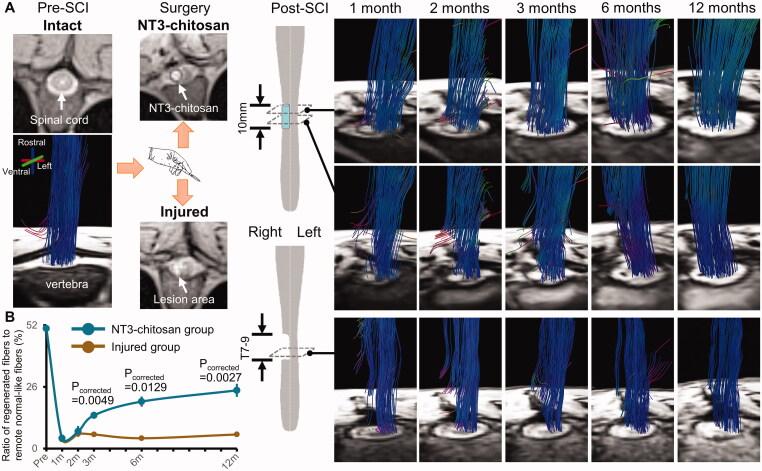
Figure 1.
Representative DTI-fibre tracking longitudinal results of injured and NT3-chitosan treated animals. Data processing just following the same methods as we previously reported [26]. (A) Reconstructed fibre bundles were superimposed on the corresponding axial PD weighted structural images. Before operation, the structure of the spinal cord exhibited good integrity, and the fibre bundles filled the whole spinal cord structure in an orderly manner. After operation, the tissue of the right thoracic cord was damaged and the structural integrity was destroyed, and the implanted NT3–chitosan scaffold could be clearly observed. The fibres (rostrocaudal orientation) gradually extended across the surgical site over time, reconnecting the rostral and caudal ends of the injured cord in the regenerative therapy animals. On the axial structural images, the material boundary gradually blurred and disappeared with the degradation of the implanted NT3-chitosan scaffold. In injured animals, no fibre bundles were present within the lesion and passed through the injured site. (B) The proportion of the regenerated fibres to the number of remote normal-like spinal cord fibres was significantly different between the two groups at 3, 6, and 12 months post-SCI (Independent t-test with Bonferroni multiple comparisons). m, months.

