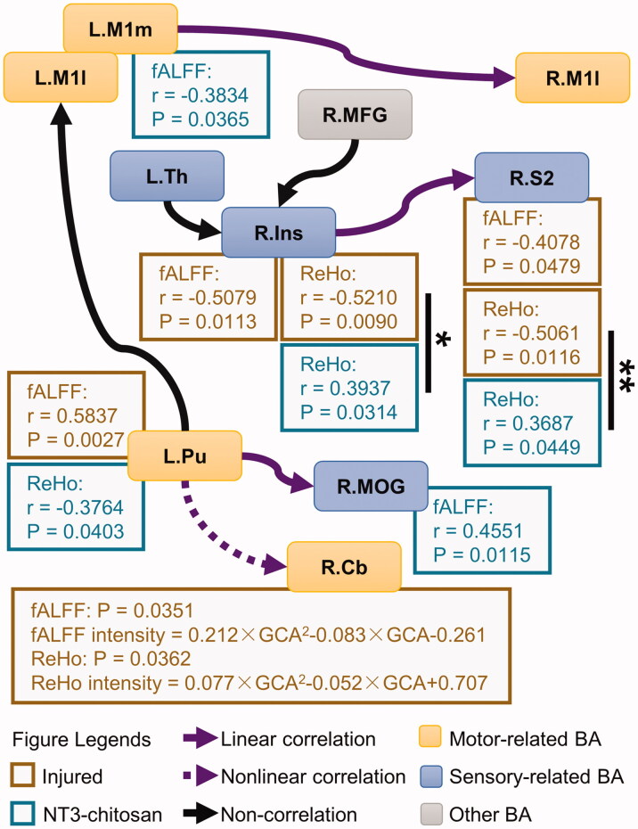
Figure 5.
The injured and NT3-chitosan-treated animals have different relationships between the intensity of information flow and the property of the spontaneous activities. Correlations between the intensity of the information flow that was affected by time × group interaction and the fALFF/ReHo values in the brain regions corresponding to these flows were shown (r and p values are given). No pronounced relationship was observed between other information flows and the properties of local brain activity. Significant differences in correlation mode between the two groups were calculated (Chow-test with Bonferroni multiple comparisons). *p < 0.05; **p < 0.01. L: left; R: right; M1l: lateral primary motor cortex; M1m: medial primary motor cortex; Th: thalamus; MFG: middle frontal gyrus; Ins: insula; S2: secondary somatosensory cortex; Pu: putamen; MOG: middle occipital gyrus; Cb: cerebellum.

