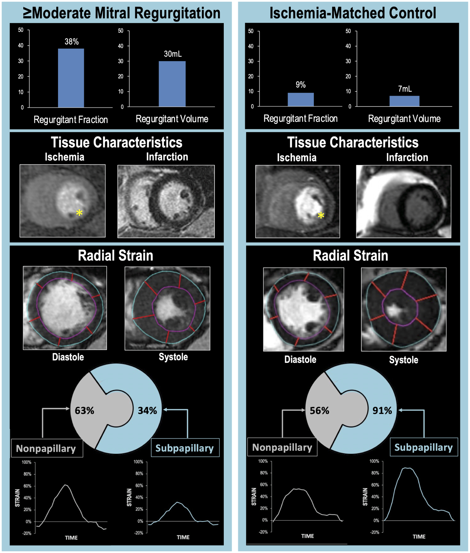
Figure 2. Typical Examples of ≥Moderate FMR in Relation to Sub-Papillary Ischemia and Strain.

Representative examples of ischemic CAD patients with (left) and without (right) ≥moderate FMR. Note that despite marked differences in quantitative severity of FMR (top), both patients manifested perfusion deficits in mitral valve adjacent (inferior/lateral) LV myocardium on stress perfusion CMR (middle left, yellow asterisks) and absence of infarction on late gadolinium enhancement CMR (middle right). Regional strain analysis (bottom) demonstrated FMR differences to parallel differential pattern and severity of impaired contractile mechanics: Despite similar strain in remote LV regions, sub-papillary radial strain (calculated as mean strain within sub-papillary segments illustrated in Figure 1A) was lower in the patient with ≥moderate FMR.
