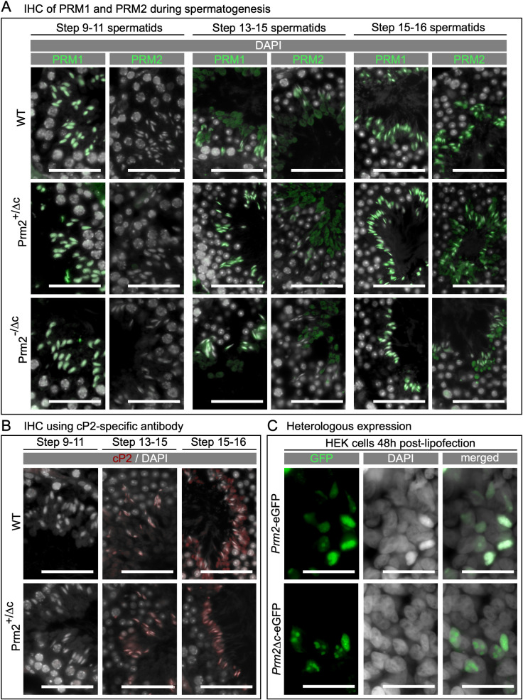
Fig 2. Localization and translation timing of PRM1, PRM2, unprocessed PRM2 and DNA condensing ability of Prm2Δc.
(A) Immunohistochemical fluorescent staining of PRM2 (WT, Prm2+/Δc) or mP2 (Prm2+/Δc, Prm2-/Δc) (green) and PRM1 (all genotypes) (green) in testis sections, counterstaining with DAPI (pseudo-colored grey). Scale bar = 50μm. (B) Immunohistochemical fluorescent staining of unprocessed PRM2 (red) using a cP2-specific antibody in testis sections, counterstained with DAPI (pseudo-colored grey). Scale bar = 50μm. C) Fluorescent images of human embryonic kidney 293 (HEK) cells 48 hours post-transfection with plasmids encoding eGFP tagged PRM2 (Prm2-eGFP) or Prm2Δc (Prm2Δc-eGFP) (green), counterstained with Hoechst (pseudo-colored grey). Scale bar = 50μm.

