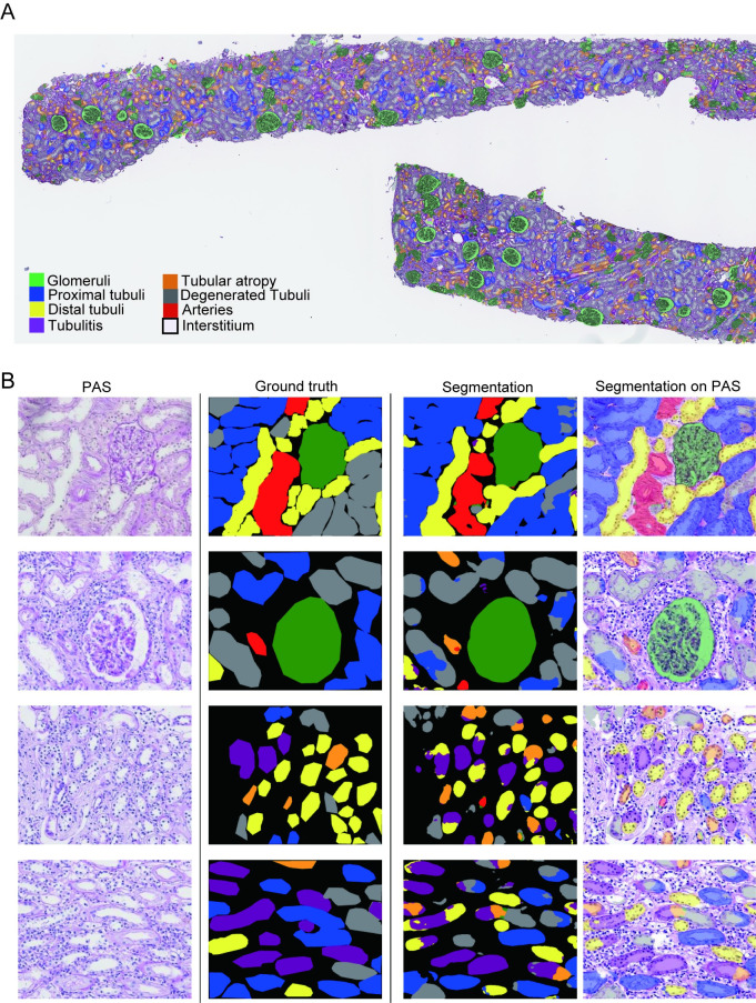
Fig 2. Representative images of ground truth and eight-class segmentation using U-Net.
(A) Whole-slide image of segmentation using U-Net in a specimen with tubulointerstitial nephritis. (B) PAS-stained slide, ground truth, and segmentation using U-Net. The top row represents a normal specimen, and the second through fourth rows represent specimens with tubulointerstitial nephritis.

