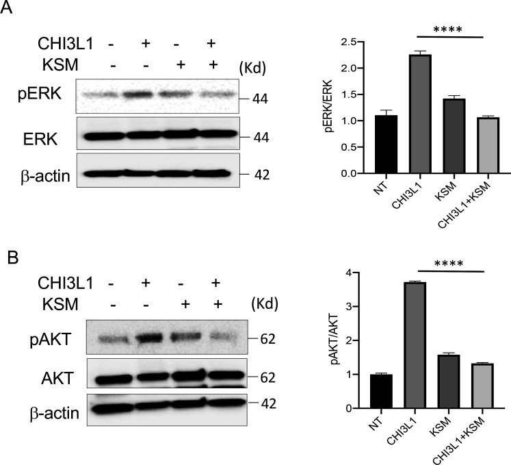
Figure 4. Kasugamycin inhibition of CHI3L1-induced signaling.
Calu-3 cells were stimulated with rhCHI3L1 (250 ng/ml) or its vehicle control for 2 hr in the presence of kasugamycin (250 ng/ml) and vehicle control (PBS). (A, B) Western blotting and densitometry analysis were then employed to evaluate the levels of activated (phosphorylated) (p) and total ERK (A) and AKT (B). The noted figure is representative of a minimum of three similar experiments. ***p<0.0001 (t-test).

