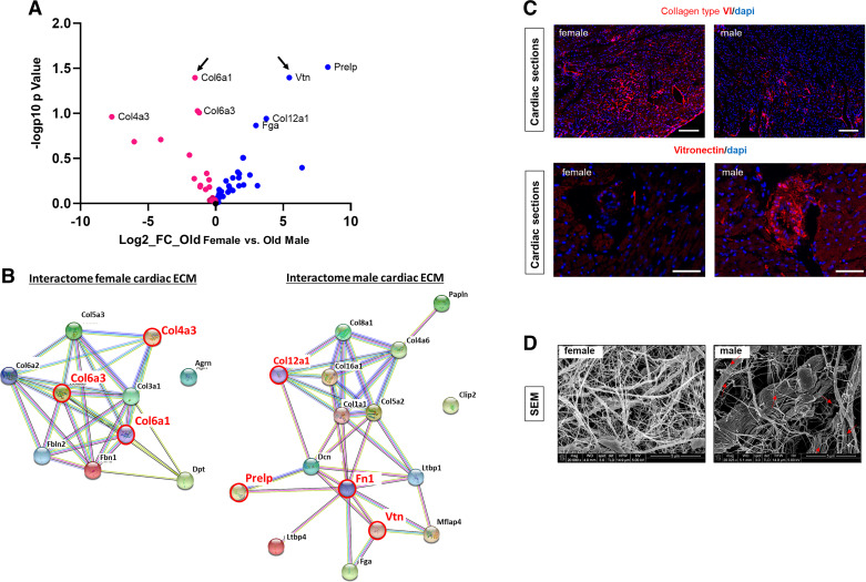
Figure 2.
Differences in extracellular matrix (ECM) composition and architecture of the aging female and male mouse hearts. A: mass spectrometry analysis. Volcano plot showing the log2 fold change (FC) against the log10 adjusted P value comparing proteins that are highly expressed in male (blue circle) vs. female (red circle) cardiac matrices. Arrows indicate proteins that have been chosen to validate mass spectrometry data by immunofluorescence staining (n = 4). B: protein-to-protein networks generated with STRING for cardiac female (left) and male (right) matrices. Red circle and red fonts depict proteins that are the most significantly different in the female and male cardiac matrices, respectively. C: representative pictures of immunostaining on cardiac cross sections from old male and female mice for collagen type VI α1 (top, Scale bar = 200 µm) and vitronectin (bottom, Scale bar = 100 μm). Nucleus counterstaining was performed with DAPI (n = 4). D: decellularized cardiac matrices prepared from old male and old female mice and visualized by scanning electron microscopy. Red arrows point to broken fibrils that are consistently found in male matrices. Scale bar = 5 μm (n = 3) N, number of animals/group..

