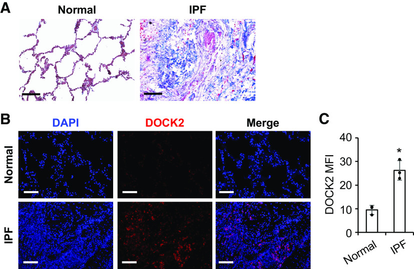
Figure 7.
DOCK2 expression was induced in lung tissues from human patients with idiopathic pulmonary fibrosis (IPF). A: representative images of Masson’s trichrome staining showed extensive collagen deposition (blue) in the lung tissues of patients with IPF (n = 3/group). B: representative IF staining showed notably increased DOCK2 expression (red) in the lung tissues from patients with IPF as compared with normal control. DAPI stains the nuclei (blue). C: the quantification and comparison of DOCK2 mean fluorescence intensity (MFI) was based on measurements from 10 representative fields per slide from each of three control and patients with IPF. *P < 0.05 compared with normal controls, as tested by a two-tailed, unpaired Student’s t test. Scale bars (black and white) represent 100 µm, images were taken at the same magnification. DOCK2, dedicator of cytokinesis 2; IF, immunofluorescence.

