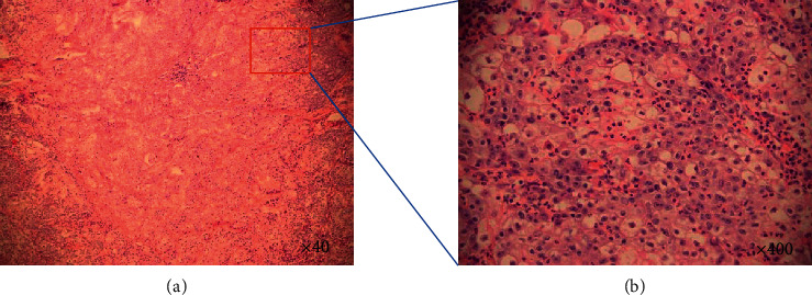
Figure 2.

Histomorphology of NUT midline carcinoma (case 3): poorly differentiated tumour cells and focal keratinization (H&E stain, ×40) (a). Higher magnification shows that most of the cells were incohesive, monotonous, atypical, and scanty-cytoplasm tumour cells with irregular ovoid hyperchromatic nuclei (H&E stain, ×400) (b).
