
Figure 4.
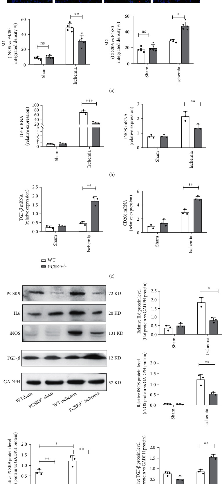
PCSK9 knockout inhibited M1 polarization and promoted M2 polarization in myocardial macrophages after infarction. (a) Representative immunofluorescence staining showing the percentages of M1 (F4/80+iNOS+CD206−) and M2 (F4/80+iNOS−CD206+) in WT/PCSK9−/− mouse myocardium after ischemia or sham. Nuclei were counterstained with DAPI. Scale bar = 50 μm, n = 5. Quantitative analysis of the percentage of M1 and M2 macrophages of (a). (b, c) q-PCR analysis of IL-6, iNOS, TGF-β, and CD206 mRNA expression in WT/PCSK9−/− mouse myocardium after ischemia or sham, n = 3. (d) Representative images of Western blots for PCSK9, IL6, iNOS, and TGF-β in WT/PCSK9−/− mouse myocardium after ischemia or sham, n = 3.