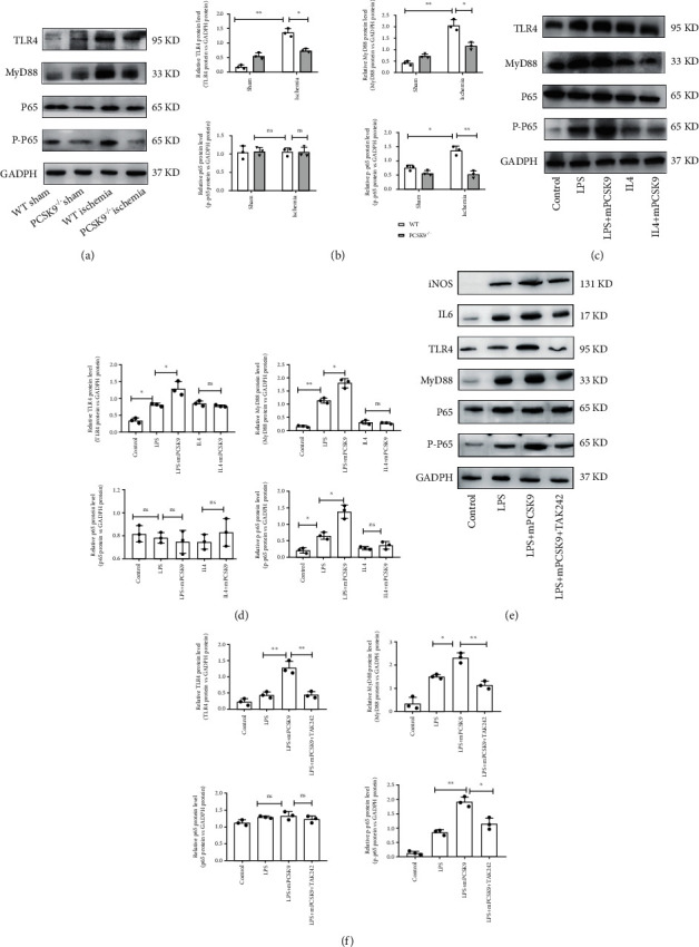
Figure 6.

PCSK9 regulated M1 macrophage polarization by targeting TLR4. Representative images of Western blots for TLR4 and downstream MyD88/NF-κB in WT/PCSK9−/− mouse myocardium after ischemia or sham, n = 3. (b) Protein levels of TLR4 and downstream MyD88/NF-κB of (a). (c) Representative images of Western blots for TLR4 and downstream MyD88/NF-κB in LPS/IL4-stimulated RAW264.7 cells after cocultivation with PCSK9 protein for 24 h, n = 3. (d) Protein levels of TLR4 and downstream MyD88/NF-κB of (c). (e)TLR4 inhibitor (TAK242) was used to analyze whether TLR4 is involved in PCSK9-regulated macrophage polarization; (f) protein levels of IL6, iNOS, TLR4, and downstream MyD88/NF-κB of (e). ∗P < 0.05; ∗∗P < 0.01; ∗∗∗P < 0.001; ns: not significant.
