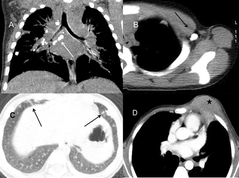
Figure 2.
Pulmonary CT of BCG infection. (A) Coronal reformatted CT image of a 1-year-old boy demonstrated multiple nodular calcification within the mediastinal and hilar lymph nodes (long arrow). The calcified nodule in the right lower lobe was also found. (B) The left axillary calcification ipsilateral to the BCG injection site was found in a 1-year-old boy (long arrow). (C) Chest CT of a 9-month-old boy showed multiple pulmonary nodules in the bilateral lower lobe beneath the pleura (long arrow). (D) A 8-month-old boy with chest wall invasion. CT demonstrated a large chest wall mass with patchy areas of low density inside, suggesting the forming of abscess (star).

