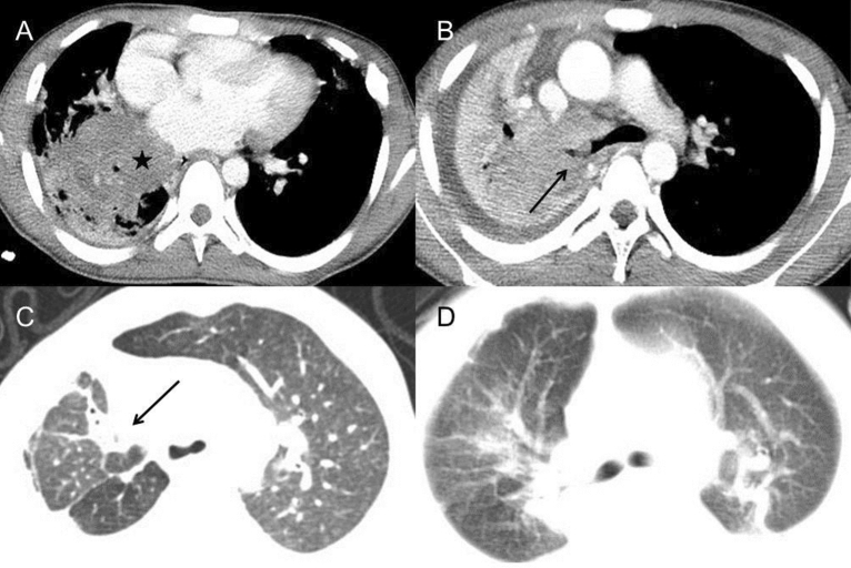
Figure 5.
Pulmonary CT of TB infection. (A,B) 16-year-old boy with TB infection. (A) Post-contrast CT showed a large mass in the right lower lobe, about 5 cm in diameter (star). Multiple small cavities were found in the adjacent area of consolidation. (B) A nodule in the right bronchus led to atelectasis of the right lung, which was identified as TB granuloma formation by bronchoscopy (long arrow). The right pleural effusion was also found. (C) In a 3-year-old boy, axial CT scan showed scarring, segmental atelectasis and volume loss in the right upper lobe (long arrow). Slight bronchiectasis could be found inside the area of consolidation. (D) Segmental consolidation in bilateral upper lobes with adjacent ground-glass opacity were found in a 2-year-old boy.

