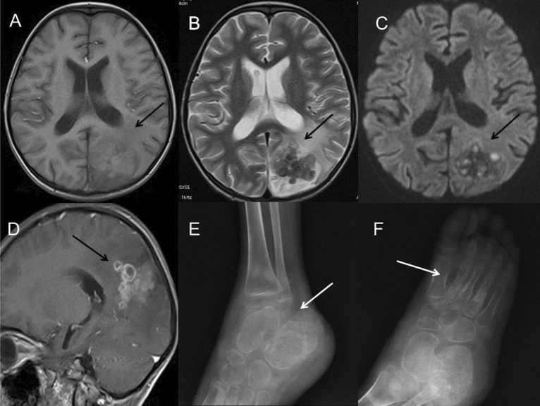
Figure 6.
A 5-year-old boy with infection of TB. (A–D) MRI showed multiple small cerebral abscesses (long arrow) in the left parietal and occipital lobes, which had high intensity on T1WI (A), low intensity on T2WI (B), high intensity on DWI sequence (C) and annular enhancement on post-contrast T1WI (D), suggesting the presence of caseous necrosis. Extensive cerebral edema could be found around the lesions. (E,F) Osteomyelitis in the right tibia and foot was found in the same patient, demonstrating local bone erosions and osteosclerosis in the lower metaphysis of the tibia, the calcaneal and the 2nd–4th metatarsal bones (long arrow). Periosteal thickening along the metatarsal bones and swelling soft tissue of the right ankle and foot were found.

