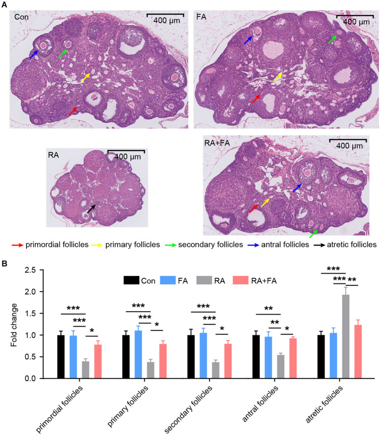
Figure 2.
Effect of folic acid (FA) pre-administration on ovarian pathological damage after radiation injury. (A) The results of hematoxylin and eosin (H&E) staining indicated that FA pre-administration relieved ovarian histopathological damage. Red arrow: primordial follicles; yellow arrow: primary follicles; green arrow: secondary follicles; blue arrow: antral follicles; black arrow: atretic follicles. Scale bar, 400 μm. (B) Relative quantification of the ovarian follicles for each group. There was no significant difference in follicle number between the Con group and FA group (p > 0.05). The numbers of primordial follicles, primary follicles, secondary follicles, and antral follicles in the RA group were significantly lower than those in the Con group. FA pre-administration significantly relieved the effect of radiation on the ovarian tissue structure and increased the number of ovarian follicles, thus enhancing ovarian reserve capacity. Con: control group, FA, FA pre-administration control group; RA, radiation exposure group; RA+FA, FA pretreatment + radiation exposure group. Error bars, standard error of mean (SEM). N = 7, *p < 0.05, **p < 0.01, and ***p < 0.001.

