Figure 4.
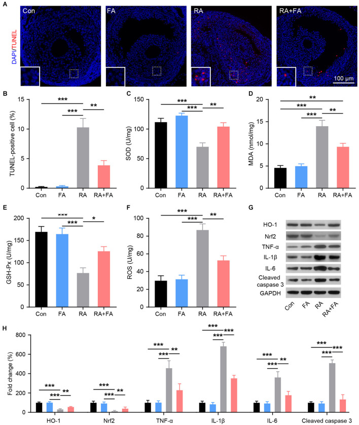
Effect of folic acid (FA) pre-administration on radiation-induced ovarian apoptosis, oxidative stress, and inflammation. (A,B) Ovarian follicle cell apoptosis was determined via TUNEL staining. Apoptotic cells are stained in red, and nuclei are stained in blue (DAPI). Scale bar, 100 μm. (C–F) Effect of FA pre-administration on the levels of SOD, MDA, GSH-Px, and ROS in ovarian homogenates. SOD, superoxide dismutase; MDA, malondialdehyde; GSH-Px, glutathione peroxidase; ROS, reactive oxygen species. Each ELISA assay was repeated three times. (G,H) Western blot was performed to detect the protein levels of HO-1, Nrf2, TNF-α, IL-1β, IL-6, and Cleaved caspase-3 in ovarian tissues from each group.in ovarian tissues from each group. HO-1, heme oxygenase 1; Nrf2, nuclear factor-erythroid 2-related factor 2. TNF-α, tumor necrosis factor-α; IL-1β, interleukin-1β; IL-6, interleukin-6; Con, control group; FA, FA pre-administration control group; RA, radiation exposure group; RA+FA, FA pretreatment + radiation exposure group. Error bars, standard error of mean (SEM). N = 8, *p < 0.05, **p < 0.01, and ***p < 0.001.

