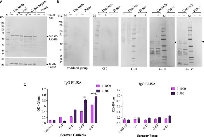
Figure 5.
In vitro and in vivo recognition of VM proteins in Leptospira cell free lysate by sera from immunized mouse groups. Pathogenic L. interrogans serovars Canicola, Lai, Copenhagni, and non-pathogenic L. biflexa serovar Patoc were grown in conditional EMJH medium which was induced with 120 mM NaCl for 4 h in log phase and unconditional EMJH medium and cells were harvested. Cell-free lysates were analyzed by 4-12% SDS-PAGE, then transferred to a nitrocellulose membrane for Western blot analysis. (A) The membrane was probed with polyclonal LA3490 antibodies (1:2,000 dilution) and LipL32 monoclonal antibody (1:10,000) which was served as loading control. (B) The other set of membrane were probed with pooled sera (1:100 dilution) collected before immunization (pre-bleed) and after challenge Group I (PBS+ adjuvant), Group II (t3490), Group III (VM Mix) and Group IV (VM Unlabeled. (A, B) Leptospira grown in EMJH medium without addition of NaCl, represented by minus (–), and Leptospira grown in EMJH medium to log phase, at which time 120 nM NaCl was added, represented by plus (+). Arrows indicate the expression of 70.29 kDa native VM proteins. (C) Anti-Leptospira immunoglobulins generated against with serovars Canicola after experimental infection of C3H/HeJ suspectable mice. Whole cell IgG ELISA were performed with pre-bleed and sera from immunized mice post-challenge. Serovar Patoc served as negative control. ***p < 0.001, ****p < 0.0001.

