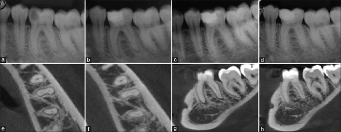
Figure 2.
Representative image of biodentine full pulpotomy in tooth #36, (a) preoperative radiograph, (b) immediate postoperative, (c) 6-month follow-up, (d) 12-month follow-up, (e) preoperative cone beam computed tomography axial section showing apical periodontitis, (f) 12-month follow-up cone beam computed tomography, (g) preoperative cone beam computed tomography sagittal section, (h) 12-month follow-up cone beam computed tomography showing healing of periapical lesion

