Figure 3.
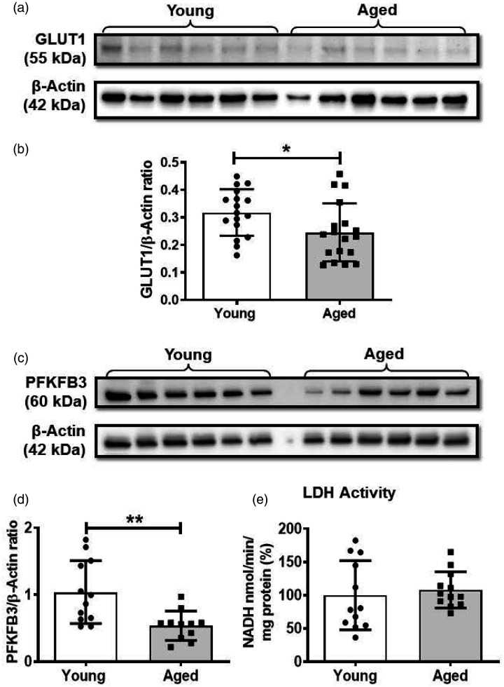
Western blot analysis revealed that the brain microvessels from the aged mice have reduced expression of glucose transporter 1 (GLUT1) and 6-phosphofructo-2-kinase/fructose-2, 6-biphosphatase 3 (PFKFB3) and unaltered lactate dehydrogenase (LDH) activity. (a). Representative western blot for GLUT1 and internal control, β-actin. (b). GLUT1 to β-actin band density ratio. (c). Western blot for PFKFB3 and internal control, β-actin. (d). PFKFB3 to β-actin band density ratio. (e). LDH activity. Data were represented as mean ± SD and analyzed by student’s t-test. P ≤ 0.05 was taken as the statistical significance. N = 17–18 mice/age group (GLUT1 expression and LDH activity), N = 12 mice/age group (PFKFB3 expression). All the original immunoblots are included in the online supplementary figures (2–5).

