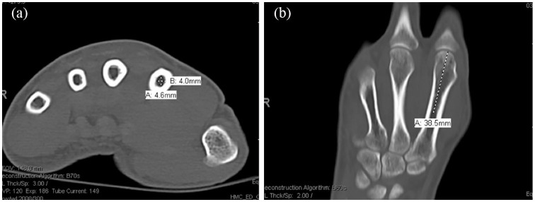
Figure 2.
Computed tomographic measurements of the metacarpal using Philips IntelliSpace PACS Enterprise. Image (a) represents an axial cut depicting the measurement performed to determine the (A) volar-dorsal and (B) radial-ulnar measurement at the narrowest part of the metacarpal shaft. Image (b) represents a coronal cut depicting the measurement performed to determine the (A) length from the metacarpal head to the narrowest point of the metacarpal shaft.

