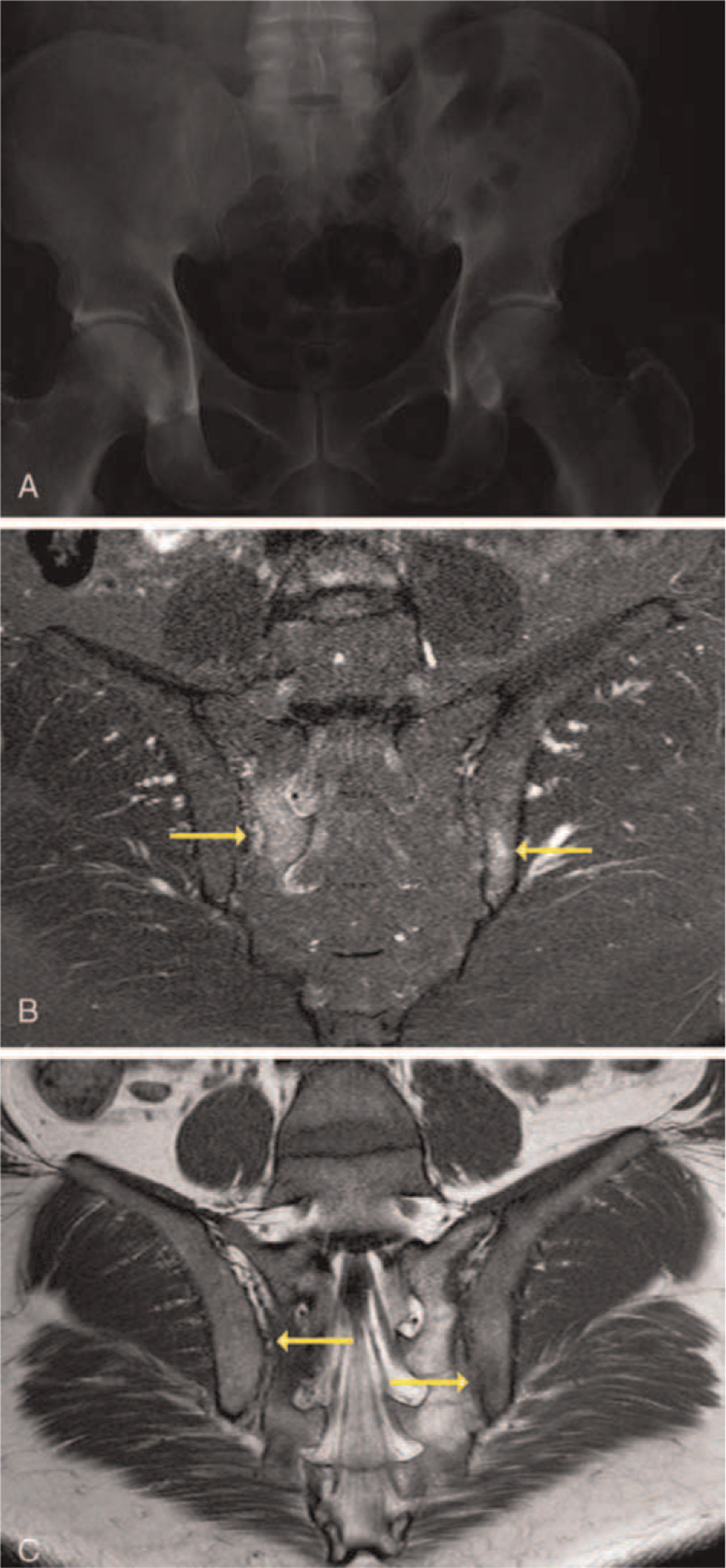
Figure 3.
MRI plays an important role in the diagnosis of nr-axSpA. Imaging plays an important role in the diagnosis of nr-axSpA. In patients with nr-axSpA, X-ray images may be completely normal as these images do not allow detection of early signs of axial inflammation.[49] The use of MRI of SIJs enables detection of osteitis (undetectable by X-ray) before radiographic sacroiliitis appears.[49–51] (A) Shown is an X-ray image of a pelvis in a 45-year-old African-American patient with a 3-month history of low back and ankle pain. Pain wakes him at night and gets worse with prolonged sitting. The patient is HLA-B27-negative with a CRP level of 8.6 mg/dL. (B) Shown is an image of a STIR image showing high intensity edema (yellow arrows) on the sacral side of the right SIJ and iliac side of the left SIJ in the same patient. (C) Shown is a T1-weighted image with hypointensity areas showing edema with erosion (yellow arrows) on the sacral side of the right SIJ and iliac side of the left SIJ in the same patient. Patient images were provided courtesy of Dr Marina Magrey. axSpA = axial spondyloarthritis, CRP = C-reactive protein, MRI = magnetic resonance imaging, nr-axSpA = nonradiographic axial spondyloarthritis, SIJ = sacroiliac joint, STIR = short tau inversion recovery.

