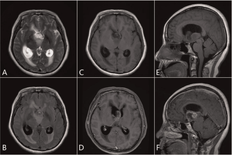
Figure 1.
The third ventricle showed isometric T1, slightly longer T2 with mild mixed signal mass shadow, cystic solid. The boundary was clear, with no definite edema signal in the adjacent brain tissue. The main body of the lesion was located in the third ventricle and spread to the right lateral ventricle. Bilateral ventricular dilatation was observed, wherein the right ventricle was more significant. A long stripe T2 signal can be seen at the edge of the lateral ventricle (interstitial brain edema). Enhanced scanning of the mass sac wall and solid components showed irregular ring enhancement, and local lace-like enhancement (A, axial T2; B, axial T2 FLAIR; C, axial T1; D, axial T1 enhancement; E, sagittal T1; F, enhanced sagittal T1).

