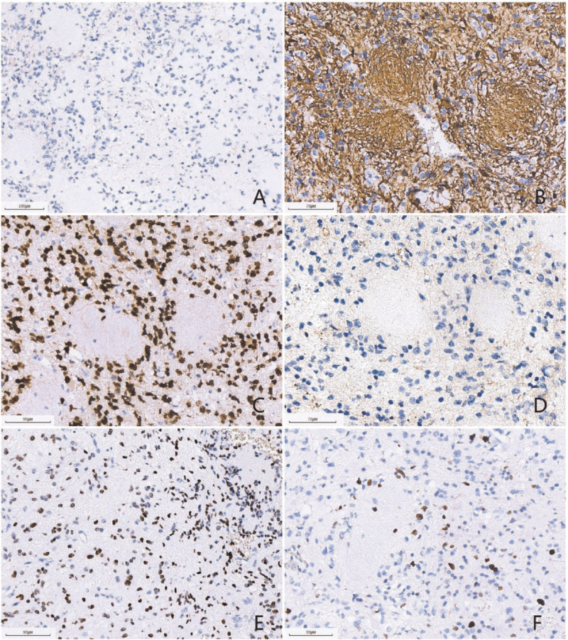
Figure 3.
(A) (scale bar: 100 μm) The tumor cells were negative for IDH1 R132H. (B) (scale bar: 70 μm) The tumor cells and “neuropil-like islands” were positive for GFAP. (C) (scale bar: 90 μm) The tumor cells were positive for Olig-2. (D) (scale bar: 70 μm) The tumor cells and “neuropil-like islands” were negative for synaptophysin. (E) (scale bar: 90 μm) The tumor cells were positive for H3-K27 M mutant protein. (F) (scale bar: 80 μm) The Ki-67 proliferation index was about 20%.

