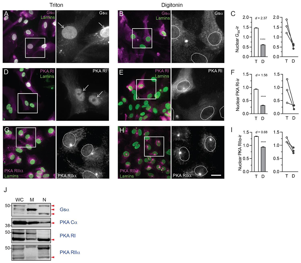
Fig. 5. Nuclear localization of β-adrenergic signaling components in mouse astrocytes.

Fluorescence photomicrographs of fixed mouse primary astrocytes permeabilized with either Triton X-100 or digitonin, followed by incubation with antibodies directed against adrenergic receptor downstream signaling components (magenta in dual color images, white in single color). All astrocytes were then permeabilized again with Triton X-100 and incubated with an antibody against lamins A and C (green) to label nuclei. Boxes indicate areas shown at higher magnification to the right. Positions of nuclei are indicated with arrows or white ovals. Images are representative of immunostaining patterns observed in n ≥ 3 cell cultures from separate litters for each of the following proteins: (A, B) Alpha subunit of the stimulatory G protein (Gsα); (D, E) PKA regulatory subunit I (PKA RI); and (G,H) PKA regulatory subunit IIα (PKA RIIα). In each pair of images, scale = 25 μm in the dual-color image, 9.25 μm in grayscale image. (C, F, I) Quantification of nuclear immunofluorescence for each target protein in Triton- and digitonin-permeabilized astrocytes. Bar graphs on the left depict mean ± s.d. nuclear immunofluorescence for each target protein from all ROIs across all biological replicates for each detergent condition. Line graphs on the right depict target protein mean nuclear immunofluorescence in each of the three biological replicates. Lines connect mean values for the two detergent conditions from the same biological replicate. Cohen’s d values are shown to indicate the size of the effect. **** P< 0.0001. (J) Immunoblotting of whole cell lysate, membrane, and nuclear proteins from primary mouse astrocytes using antibodies against Gsα, PKA catalytic subunit (PKA Cα), PKA RI, and PKA RIIα. Red arrowheads indicate positions of predicted molecular weights for the protein of interest. All images are representative of immunostaining patterns observed in n = 3 cell cultures from separate litters.
