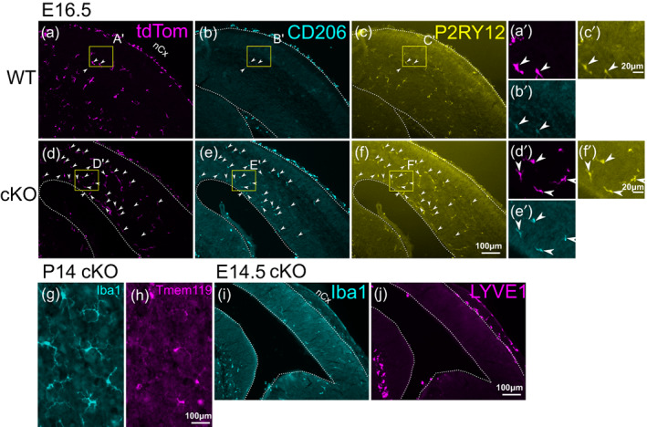
FIGURE 5.

Analysis of marker expression in Iba1+ cells in Sall1 mutant brains. (a–f) Triple immunostaining of tdTomato (a,d) CD206 (b,e) and P2RY12 (c,f) in wild type (Lyve1 Cre/+ ; tdTomato reporter; Sall1 wt/wt ) and Sall1 cKO (Lyve1 Cre/+ ; tdTomato reporter; Sall1 flox/flox ) embryos at E16.5. Arrowheads indicate cells that express all three markers. Most of the persistent CD206+ cells in the parenchyma of the cKO brains are tdTomato+ and also express the microglia marker, P2RY12. (g,h) Double immunostaining of Iba1 and Tmem119 in Sall1 knockout cortex at P14, showing that parenchymal cells that express Iba1 still express the microglia marker, Tmem119. (i,j) Double immunostaining of Iba1 (i) and LYVE1 (j) in Sall1 cKO brain at E14.5 showing that mutant microglia do not express the BAM marker, LYVE1. For a–f, a high‐magnification image is shown (e.g., a' for a). The location of each high‐magnification image is shown as a yellow box in each panel.
