FIGURE 7.

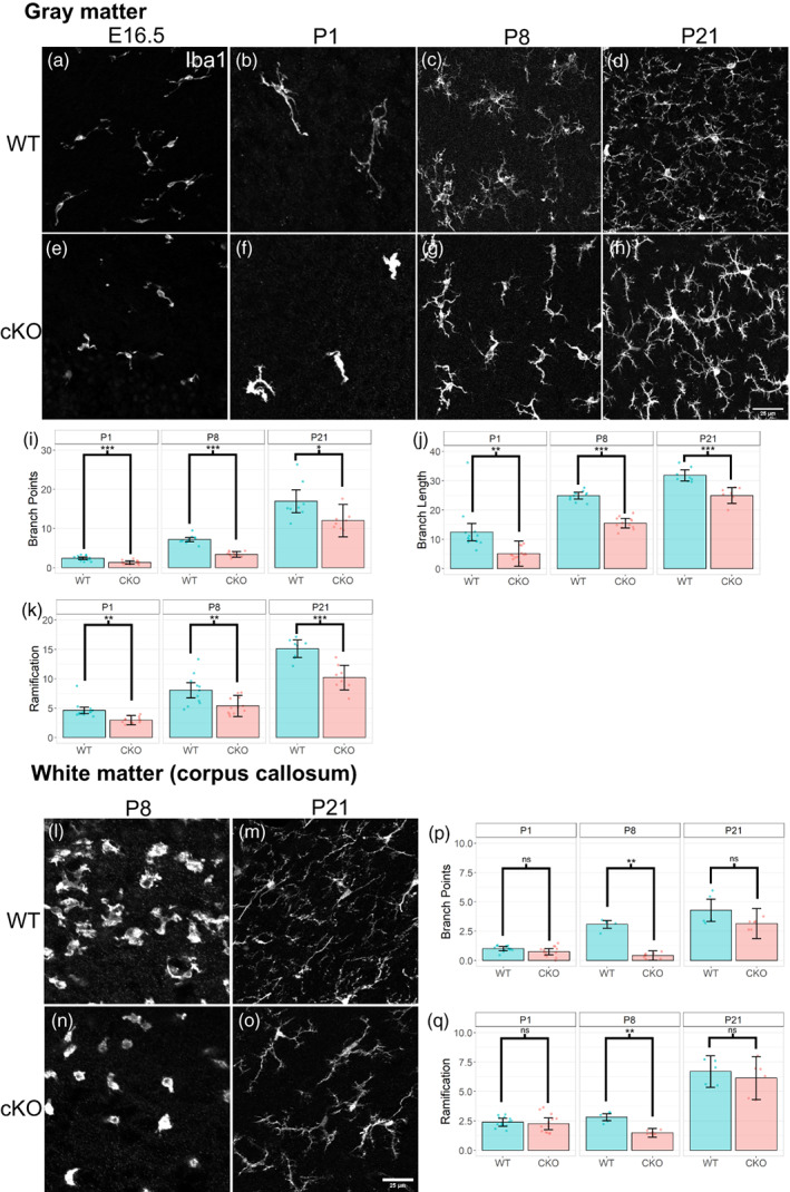
Altered microglia morphology in Sall1 mutant brains. (a–h) Immunostaining of Iba1 in the gray matter of the somatosensory cortex of wild type (a–d) and Sall1 cKO (e–h) mice at E16.5 (a,e), P1 (b,f), P8 (c,g) and P21 (d,h). Sall1 mutant microglia show less ramified morphology and less coverage of space. All images were taken with a confocal microscope and represents a z‐stack of 8 slices (0.49 μm/slice). (i–k) Quantitative analysis of the morphology gray matter microglia using the 3DMorph program. Numbers of branch points (i), branch length (j) and ramification indexes (k) are compared between wild type (blue bars) and Sall1 cKO (pink bars) primary somatosensory cortex. (l–o) Immunostaining of Iba1 in the white matter of the somatosensory cortex of wild type (l,m) and Sall1 cKO (n,o) mice at P8 (l,n) and P21 (n,o). Sall1 mutant microglia show less ramified morphology and less coverage of space. All images were taken with a confocal microscope and represents a z‐stack of 8 slices (0.49 μm/slice). (p,q) Quantitative analysis of the morphology of white matter microglia using the 3DMorph program. Numbers of branch points (p) and ramification indexes (q) are compared between wild type (blue bars) and Sall1 cKO (pink bars) primary somatosensory cortex.
