
Fig. 6. Heterotypic tissue collisions.
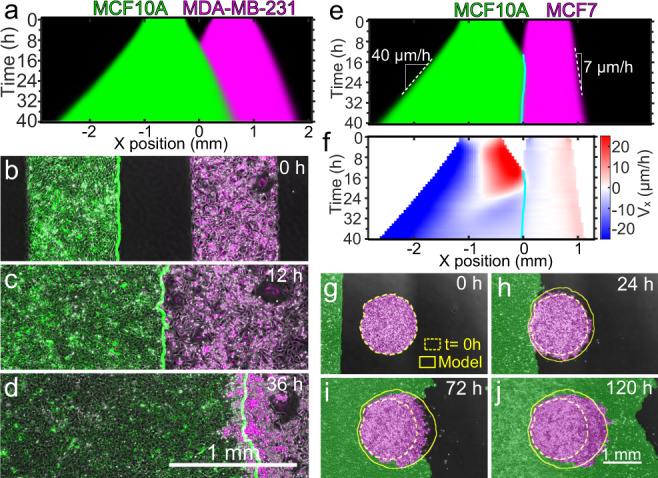
a Average kymograph of segmented fluorescence for collisions (n = 12) between rectangular MCF10A and MDA-MB-231 tissues of the same size and cell density. The MCF10A tissue displaces the MDA-MB-231 tissue. b–d Snapshots of the co-culture at the initial configuration (b), the collision time t = 12 h (c), and 24 h after collision, i.e., t = 36 h (d). The green line indicates the edge of MCF10A tissue. Micrographs are representative of 11 independent tissue collisions with similar results. e, f Average kymographs of segmented fluorescence (e) and velocity vx (f) for collisions (n = 11) between rectangular MCF10A and MCF7 monolayers of the same size and cell density. The cyan line indicates the tissue boundary. g–j Snapshots of the initial configuration (g), collision onset (h), partial engulfment (i), and full engulfment (j) of a circular MCF7 (magenta) tissue by a rectangular MCF10A (green) tissue. Simulations (yellow outlines) confirm that a difference in expansion speed is sufficient to predict the engulfment process. Engulfment repeated with similar results in two independent experiments. See Supplementary Videos 13–16.