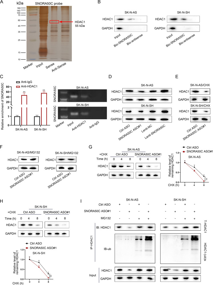
Fig. 6. SNORA50C binds to and stabilizes HDAC1 protein.
A Silver staining displayed proteins pulled down by SNORA50C and its antisense RNA. B, C RNA pull-down and RIP assays detected the binding of HDAC1 and SNORA50C in SK-N-AS and SK-N-SH cells. (Student’s t-test) **P < 0.01. Error bars indicate mean ± SD. (N = 3). D Western blot analyzed the level of HDAC1 protein in SK-N-AS and SK-N-SH cells with SNORA50C silence or overexpression. E, F Western blot showed the expression of HDAC1 protein in SNORA50C-depleted SK-N-AS and SK-N-SH cells with or without the protein synthesis inhibitor CHX or the proteasome inhibitor MG132. G, H Western blot detected HDAC1 levels in SK-N-AS and SK-N-SH cells transfected with Ctrl ASO or SNORA50C ASO#1 followed by CHX treatment at the indicated time points (left panel). Quantification of western blot results (right panel). (Student’s t-test) **P < 0.01. Error bars indicate mean ± SD. (N = 3). I SK-N-AS and SK-N-SH cells were transfected with SNORA50C ASO#1 and treated with MG132, and then cells were subjected to western blot analysis using anti-HDAC1 or anti-ubiquitin antibody.

