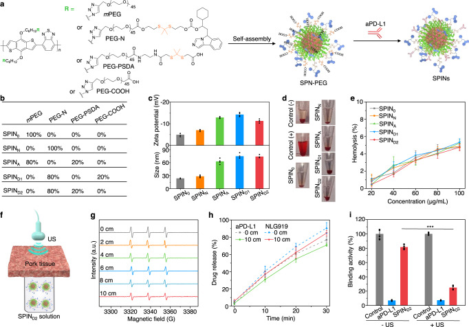
Fig. 3. Synthesis, characterization, and sonodynamic activation of SPINs.
a Chemical structures of amphiphilic semiconducting polymeric modulators and schematic illustration of their self-assembly and surface modification to form SPINs. b The molar ratios of each component in different SPINs. c Zeta potentials and hydrodynamic sizes of different SPINs in 1× PBS buffer (pH = 7.4) (n = 4). d Photographs of erythrocytes after incubation with 1× PBS buffer (negative control), 1% Triton X-100 (positive control), and 1× PBS buffer containing SPINs at the concentration of 100 µg/mL for 2 h, followed by centrifugation. e Hemolysis percentages of erythrocytes after incubation with SPINs at different concentrations for 2 h (n = 4). f Schematic illustration of US irradiation of SPIND2 solutions covered with a pork tissue. g ESR spectra of 1O2 for SPIND2 (20 µg/mL) after US irradiation (1.2 W/cm2, 3 min) without or with coverage of pork tissues at different thicknesses. h Release profiles of aPD-L1 and NLG919 from SPIND2 (40 µg/mL) after US irradiation for different time (n = 4). i PD-L1/PD-1 binding activity assay after treatment with free aPD-L1 or SPIND2 (40 µg/mL) with or without US irradiation (n = 4). SPIND2 – US versus SPIND2 + US: P < 0.0001. Statistical significance was calculated via a two-tailed Student’s t test. ***P < 0.001. In (g–i), the power intensity of US irradiation was 1.2 W/cm2 (1.0 MHz, 50% duty cycle). Data are presented as mean values ± SD. Source data are provided as a Source Data file.

