Fig. 7.
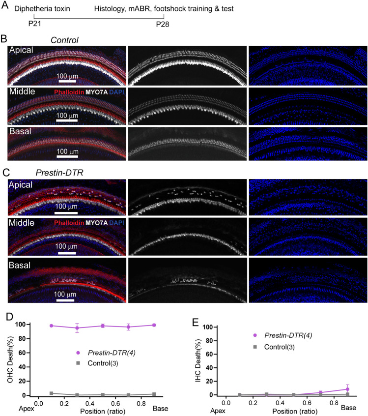
Cochlear OHC loss in DT-injected Prestin-DTR mice. A Schedule of DT injection and related tests. B Immunostaining image showing hair cell status in a control mouse at P28. Enlarged images show survival of OHCs in apical, middle, and basal locations. The cochlea is labeled with MYO7A antibody (white), phalloidin (red), and DAPI (blue). Note that most OHCs are lost but IHCs are not. Scale bar, 100 μm. C Immunostaining image showing hair cell status in a Prestin-DTR mouse at P28. The staining protocol and display conditions are as in (B). Scale bar, 100 μm. D, E Percentage loss of OHCs (D) and IHCs (E) at locations along the cochlea coil of P28 Prestin-DTR and control mice. N numbers are shown in panels.

