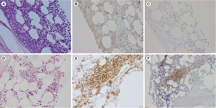
Fig. 1.
BM biopsy section and IHC staining results at diagnosis showing an increase in blasts and scattered mast cells (A, B, C) and evident mast cell aggregates post induction (D, E, F). (A, D) Hematoxylin-eosin stain (×400). (B, E) CD117 positivity in mast cells (×400). (C, F) Aberrant CD25 expression in atypical mast cells (×400).
Abbreviations: BM, bone marrow; IHC, immunohistochemical.

