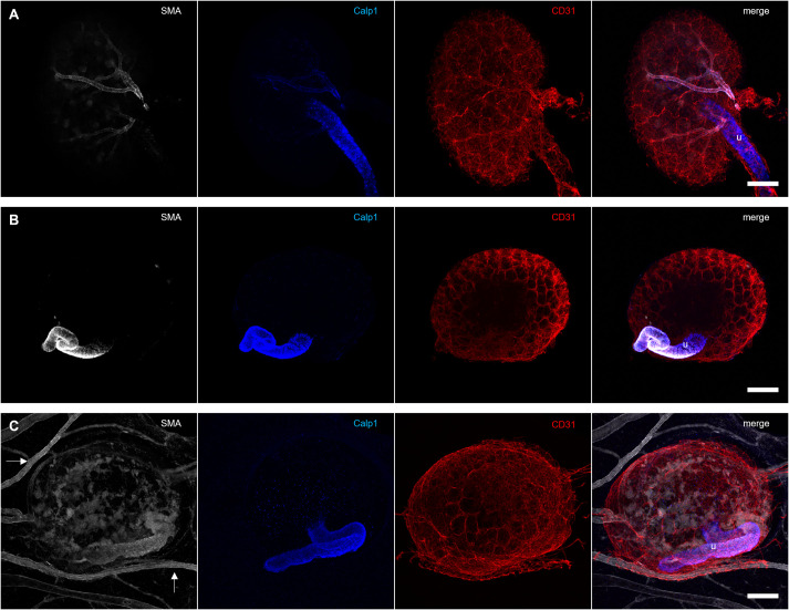
Fig. 4.
Vascular smooth muscle cells are lost in grafted kidneys. (A) Isolated E14.5 kidneys show strong smooth muscle actin (SMA) and weak Calponin 1 (Calp1) staining around the interlobular arteries. The ureter (u) stains strongly for Calponin 1. (B) E14.5 kidneys cultured for 3 days in type 1 collagen display a positive smooth muscle actin and Calponin 1 staining around the ureter, but none of the CD31-stained blood vessels are surrounded by smooth muscle cells. (C) Smooth muscle actin was detected in the galline vessels and around the ureter (u) of grafted E14.5 kidneys. Within the graft, patches with diffuse smooth muscle actin staining were visible. However, there was no alignment of smooth muscle cells around the murine CD31-positive capillaries visible. Calponin 1 was detected only in the ureter of the graft. Scale bars: 200 µm.

