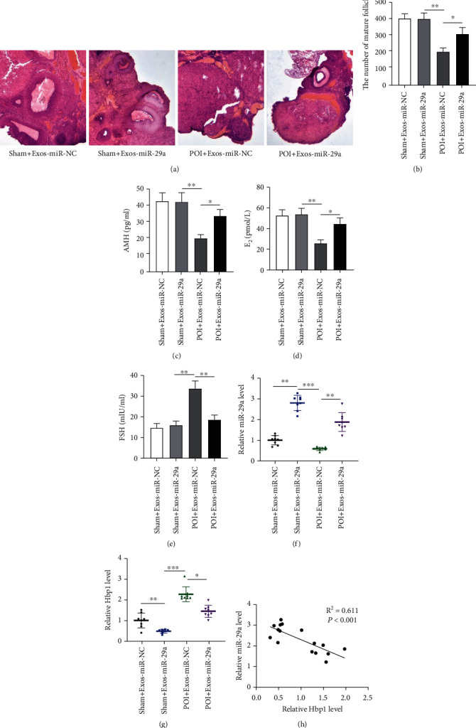
Figure 7.

The exosome carrying miR-29a improves the ovarian function in vivo. (a) H&E staining was performed to examine the histopathological changes of ovarian tissues in the sham+Exos-miR-NC group, the sham+Exos-miR-29a group, the POI+Exos-miR-NC group, and POI+Exos-miR-29a group. (b) Quantification on follicle count in different groups. (c–e) The levels of AMH, E2, and FSH in indicated groups. (f, g) The levels of miR-29a and HBP1 in ovarian tissues. (h) The correlation of miR-29a and HBP1. ∗p < 0.05, ∗∗p < 0.01, and ∗∗∗p < 0.001.
