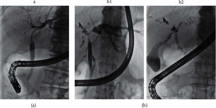
Figure 1.

Placement of each stent. (a) The IS case. A patient with Bismuth-Corlette type IIIa PHCC for whom right hepatectomy was the expected surgical operation. Because B2, B3, and B4 confluence almost simultaneously, the IS (7 Fr 9 cm deep angle) was placed in the left bile duct. (b) The FCSEMS case. A patient with Bismuth-Corlette type IIIa PHCC for whom right hepatectomy was the expected surgical operation. The cholangiogram shows that the confluence of B4 is more than 5 mm from the upper end of the stenosis (b1); FCSEMS (6 mm × 4 cm) was placed such that B4 was not obstructed (b2). IS, inside stent; FCSEMS, fully covered self-expandable metallic stent; PHCC, perihilar cholangiocarcinoma; B2, left lateral superior segmental bile duct; B3, left lateral inferior segmental bile duct; B4, left medial segmental bile duct.
