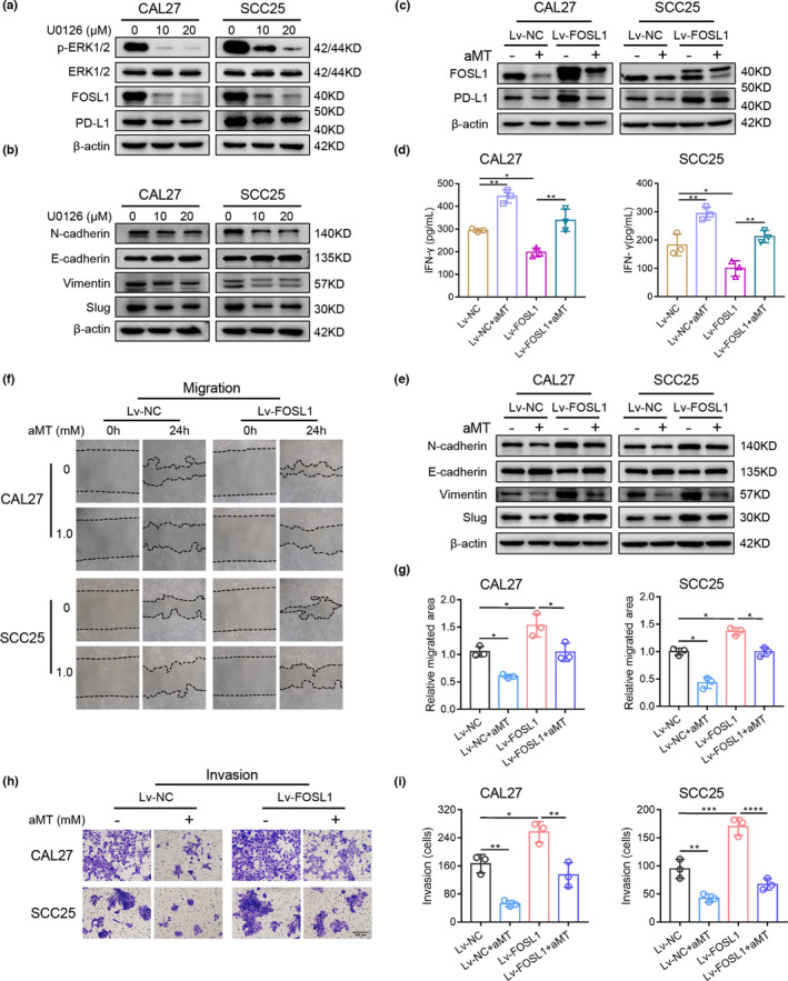
FIGURE 5.

The ERK1/2/FOSL1 Pathway is involved in melatonin‐induced inhibition of EMT and PD‐L1 expression in HNSCC cells. (A) Western blot analysis of p‐ERK1/2, ERK1/2, FOSL1, and PD‐L1 in CAL27 and SCC25 cells treated with 10 and 20 μM MEK inhibitor (U0126) for 24 h. (B) Western blot analysis of EMT‐related markers, E‐cadherin, N‐cadherin, vimentin, and Slug in CAL27 and SCC25 cells treated with 10 or 20 μM U0126 for 24 h. β‐Actin was used as the loading control. (C) CAL27 and SCC25 cells were transfected with an empty carrier lentivirus (negative control, Lv‐NC) or a FOSL1 overexpression lentivirus (Lv‐FOSL1). Lv‐NC or Lv‐FOSL1 cells were treated with or without melatonin (1 mM) for 48 h, then the protein expression levels of FOSL1 and PD‐L1 were determined by western blotting. (D) FOSL1‐overexpressed CAL27 and SCC25 cells were treated as described in Figure 5C before coculturing with pre‐activated T cells from healthy donors. After 48 h of coculture, supernatants were collected to measure IFN‐γ levels using ELISA. One‐way ANOVA. (E) FOSL1‐overexpressed CAL27 and SCC25 cells were treated with or without melatonin (1 mM) for 48 h, then the expression levels of the EMT‐related markers were determined by western blot analysis. (F–I) Wound healing assay and Matrigel invasion assay were performed and analyzed to determine the migration and invasion abilities of FOSL1‐overexpressed CAL27 and SCC25 cells pretreated with melatonin (1 mM) for 24 h. One‐way ANOVA. Scale bar, 100 μm. *, p < 0.05; **, p < 0.01; ***, p < 0.001; ****, p < 0.0001. Independent experiments were performed in triplicate. Values are represented as means ± SD
