
FIGURE 1.
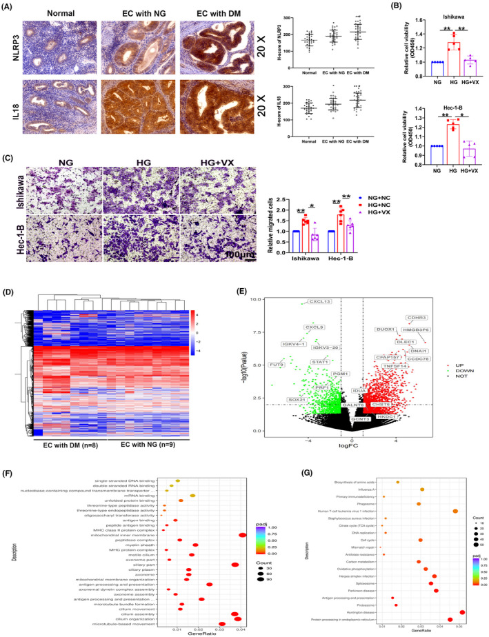
Pyroptosis is involved in EC progression. (A) Representative images of immunohistochemical staining for NLRP3 and IL18 in normal endometrial tissues (normal), tumor tissues from patients with EC presenting normal blood glucose levels (EC with NG), and tumor tissues from patients with EC complicated with diabetes mellitus (EC with DM), (n = 30). (B, C) CCK‐8 and transwell assays were used to detect the proliferation and migration capacity of Ishikawa and Hec‐1‐B cells treated with the pyroptosis inhibitor VX‐765 (VX), (n = 5–6). (D) RNA sequencing data from EC tissues from patients (n = 17). (E) Volcano map of RNA expression in EC tissues. (F‐G) Gene Ontology (GO) and Kyoto Gene and Genomic Encyclopedia (KEGG) analyses of differentially expressed genes. All values are presented as the means ± SD, * P < 0.05 and ** P < 0.01 compared with normal, # P < 0.05 compared with EC with NG (A), * P < 0.05 and ** P < 0.01 (B, C). EC, endometrial cancer; HG, high glucose; NG, normal glucose