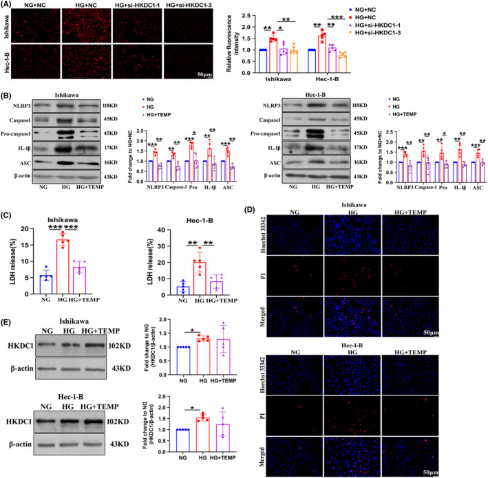
FIGURE 4.

TEMPOL prevents the HG‐induced pyroptosis of Ishikawa and Hec‐1‐B cells. (A) Mito‐SOX Red (1 µM), a mitochondrial superoxide indicator, was used to detect mitochondrial‐derived ROS production in EC cells (n = 6). (B) The expression of pyroptosis‐related proteins in EC cells treated with TEMPOL (n = 5). (C) LDH release from EC cells treated with TEMPOL (n = 5). (D) TEMPOL reduced the PI‐positive staining (red) induced by HG in EC cells (n = 3). Scale bars = 50 µm. (E) The expression of HKDC1 was determined in EC cell lines after exposure to HG for 24 h. All values are presented as the means ± SD, * P < 0.05, ** P < 0.01, and *** P < 0.001. EC, endometrial cancer; HG, high glucose; NC, negative control; NG, normal glucose; si‐HKDC1, small interfering RNA targeting HKDC1; TEMP, TEMPOL
