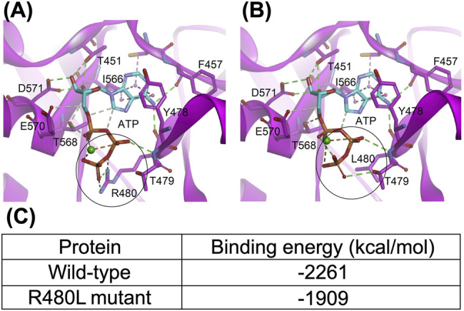
FIGURE 6.

Structure of the CNNM-PRL complex (PDB code: 5LXQ). (A) and (B), the wild-type and R480L mutant CBS module of CNNM2, respectively. The dashed lines represent the hydrogen bond (green), electrostatic (orange), and hydrophobic (pink) interactions. The metal ion is shown as a green sphere. The black circle highlights the mutation site; (C) indicates that this mutation in the CBS module results in significant impairment of the binding ability with MgATP.
