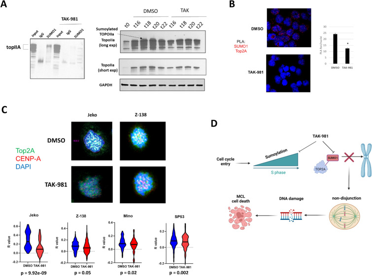
Fig. 7.
Inhibition of sumoylation with TAK-981 results in loss of centromeric localization of TopIIA in MCL cells A, left. Jeko cell were synchronized with Palbociclib (500 nM, 24 h) followed by drug washout and treatment with nocodazole (50 ng/mL) either in the presence of DMSO or TAK-981 (100 nM) for 24 h. Lysates were immunoprecipitated with antibodies directed towards SUMO1 and blotted for TopIIA. A, right. Jeko cells were synchronized with palbociclib (500 nM) and treated with either DMSO or TAK-981 (100 nM). Lysates were prepared at the indicated time points after washout and blotted for TopIIA. B, Jeko cells prepared as above were washed out of drug and fixed after 15 min. PLA for SUMO1 and topoIIA was performed. Number of individual PLA signals was quantified (n = 90 cells per condition). C Jeko (n = 88), Z-138 (n = 270), Mino (n = 80) and SP53 (n = 214) cells were prepared as above and microscopy was performed for TopoIIA and CENP-A to mark centromeric regions. The extent of colocalization of TopoIIA and CENP-A in either DMSO or TAK-981 treated cells was determined by cellsense (see “Methods” section n = 2 independent experiments per cell line). D Diagram of mechanism of desumoylation mediated cell death in MCL cells. Created on Biorender.com

