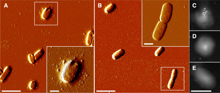
Fig. 3.
Atomic force microscopy (AFM) and agglutination tests with bacteria from static cultures. A AFM imaging of wild-type E. coli + fimA plasmid grown in a static liquid culture. A E. coli cell with type 1 fimbriae is shown in the inset. B AFM imaging of the ΔfimA1::L3S2P56 mutant E. coli + fimA+ plasmid grown in a static liquid culture. An E. coli cell with flagella is shown as an inset. C Yeast agglutination assay of wild-type E. coli + fimA+ plasmid grown in a static liquid culture. D Yeast agglutination assay in the presence of α-methyl-mannoside of of wild-type E. coli + fimA+ plasmid grown in a static liquid culture in the presence of α-methyl-mannoside. E Yeast agglutination assay of the ΔfimA1::L3S2P56 mutant E. coli + fimA+ plasmid grown in a static liquid culture. Scale bars: A, B 4 μm. A, B Insets 1 μm. C–E 2 cm

