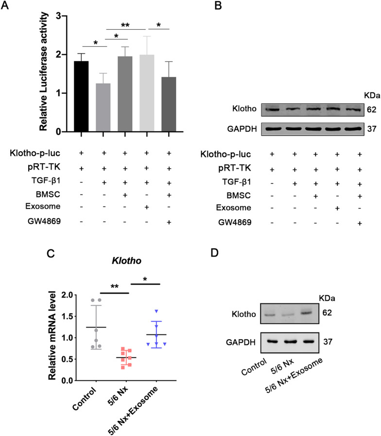
Fig. 5.
Effect of exosome transplantation on klotho activation and expression. A Fluorescent report plasmid containing klotho promoter and pRL-TK were co-transfected into 293 T cells. The transfected cells were randomly divided into five groups: control group (PBS), TGF-β1 group (1 ng/μL TGF-β1), BMSCs group (1 ng/μL TGF-β1+co-cultured with 5 × 10⁴ well BMSCs), exosomes (1 ng/μL TGF-β1+0.4 mg exosomes), and exosome inhibitor group (1 ng/μL TGF-β1+co-cultured with 5 × 10⁴ well BMSCs+GW4869). Then the firefly and Renilla luciferase activities were measured using the dual luciferase reporter gene assay. Data are shown as mean ± SD (n = 3). *p < 0.05, **p < 0.01. The protein expression of klotho was also evaluated using western blotting B, C qRT-PCR were performed to detect the mRNA expression of klotho. mRNA expression levels were normalized to that of GAPDH. Quantitative data (n = 6–7) are provided as the mean ± SD. *p < 0.05, **p < 0.01. D Expression of klotho protein in the control, 5/6 Nx, and exosome-treated 5/6 Nx rats was evaluated using western blotting

