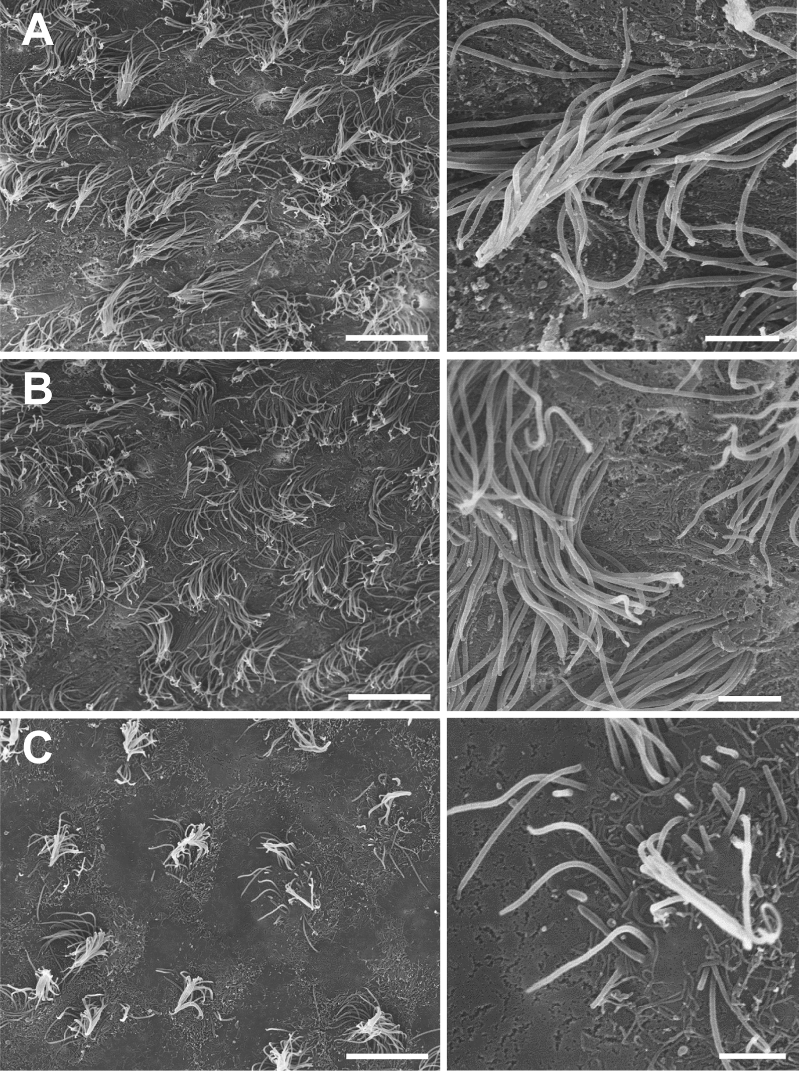
Fig. 2.

The ventricular wall presents cilia-devoid patches in LV-proximal GBM groups. Scanning electron microscopy images of the ependymal layer of mice injected with LV-proximal vehicle (A), LV-Distal GBM (B), and LV-Proximal GBM (C) (n = 3 per group). There is a progressive decrease in the number and uniformity of ependymal cilia that correlates with GBM-LV distance. Scale bars = 10 µm for larger image, 2 µm for zoomed photos. A Scanning electron microscopy images of LV-proximal vehicle injected mice showing an ependymal cell surface covered by healthy and directional cilia. B Scanning electron microscopy images of LV-distal GBM injected mice showing an ependymal cell surface covered by healthy cilia. C Scanning electron microscopy images of LV-proximal GBM injected mice showing an ependymal cell surface with unhealthy and short cilia. Scale bars = 10 µm for larger image, 2 µm for zoomed photos
