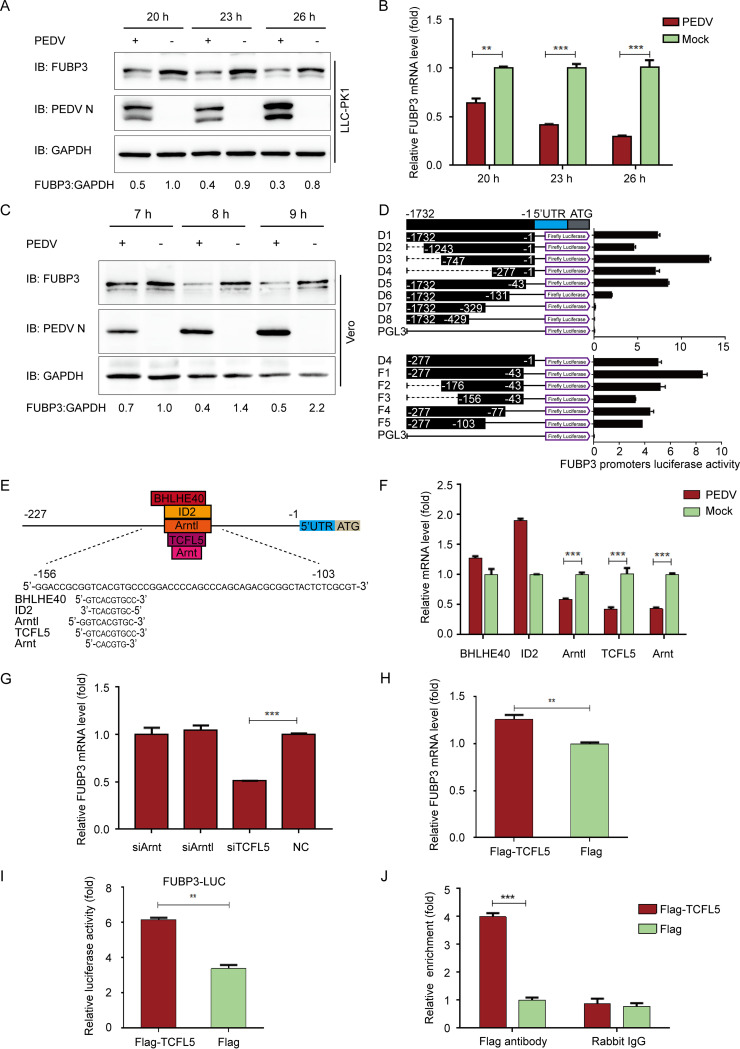
FIG 1.
PEDV infection reduces the expression of FUBP3 by the transcription factor of TCFL5 in LLC-PK1 cells. (A and B) After the infection of PEDV (MOI = 1), we harvested the cells at specific time points. Endogenous FUBP3 expression was determined by performing qRT-PCR and WB; DAPDH was used as the endogenous reference. (C) After the infection of PEDV (MOI = 1), we harvested the Vero cells at specific time points. Endogenous FUBP3 expression was determined by WB. (D) 293T cells were transfected with the truncated FUBP3 promoter constructs (D1 to D8, and F1 to F5) and Renilla luciferase reporter vector (pRL-TK-Luc) (D1 to D8, F1 to F5). The luciferase activity was measured in lysates. (E) The JASPAR vertebrate database (http://jaspar.genereg.net) was used to determine the cis-acting elements of FUBP3. (F) Arntl, BHLHE40, TCFL5, ID2, and Arnt mRNA levels in PEDV infected-PK1 cells were detected through qRT-PCR. (G) Arntl siRNA, TCFL5 siRNA, or Arnt siRNA was transfected into LLC-PK1 cells, and FUBP3 transcription was determined by performing qRT-PCR. (H) The plasmid that encoded Flag-TCFL5 was transfected in LLC-PK1 cells for a whole day, and qRT-PCR was conducted for analyzing the lysate. (I) 293T cells were subjected to transfection with FUBP3 promoter-driven luciferase vector, plasmids encoding Flag-TCFL5, and pRL-TK-Luc vector for 24 h. By analyzing the luciferase activity, the cells were collected. (J) An empty vector or a Flag-TCFL5 plasmid was transfected into LLC-PK1 cells for a day, and later, the cells were collected to conduct a ChIP assay. The normal rabbit IgG or anti-Flag antibody was adopted for precipitating chromatin-bound TCFL5. The findings are denoted as the mean ±SD from triplicate samples; *, P < 0.05; **, P < 0.01; ***, P < 0.001 (two-tailed Student's t test).

