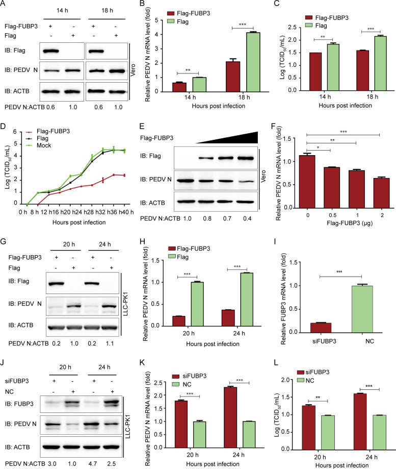
FIG 2.
The antiviral effect of FUBP3 against PEDV. (A and B) Vero cells were transfected with plasmid encoding Flag-FUBP3 and infected with PEDV (MOI = 0.01) and harvested at indicated times. qRT-PCR and WB were carried out to analyze cell lysates; ACTB served as the sample loading control. (C and D) Flag-FUBP3 was transfected into Vero cells, with the cells being infected with PEDV (MOI = 0.01). The culture supernatant was harvested at specific time points, with the viral titers being measured as TCID50 (E and F). The enhancing contents of a vector expressing Flag-FUBP3 (wedge) with the infection of PEDV (MOI = 0.01). qRT-PCR and a WB were conducted to analyze cell lysates. (G and H) LLC-PK1 cells were transfected with the plasmid that encoded Flag-FUBP3. PEDV (MOI = 1) was injected into the cells, which were collected at specific time points. Later, qRT-PCR and WB were conducted to analyze the cell lysates. (I) The knockdown efficiency of the FUBP3 siRNA in LLC-PK1 cells was analyzed by real-time PCR. (J, K, and L) LLC-PK1 cells were transfected using FUBP3 siRNA or negative control siRNA, followed by infection using PEDV (MOI = 1) and harvest the cells and culture supernatant at specific time points. PEDV N was examined by performing qRT-PCR and WB, and the viral titer was measured as TCID50. The obtained data are indicated as means ± SD of triplicate samples; *, P < 0.05; **, P < 0.01; ***, P < 0.001 (two-tailed Student's t test).

