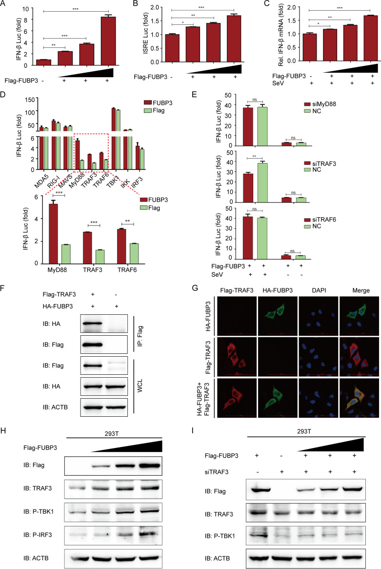
FIG 6.
FUBP3 activated the IFN signaling pathway by increasing TRAF3 expression. (A and B). The ISRE or IFNB luciferase reporter was cotransfected into HEK 293T cells with the Flag-FUBP3 expression vector (wedge) at increasing doses. (C) The lysates of HEK 293T cells were subjected to transfection with the Flag-FUBP3 expression vector (wedge), and subsequently, the Sendai virus (SeV) was used to infect cells and then conduct the luciferase assay. (D) The plasmids that encoded the FUBP3 and IFNB luciferase reporter were transfected into HEK 293 T cells with plasmids encoding MDA5, RIG-I, MAVS, MyD88, TRAF3, TRAF6, TBK1, IKK, or IRF3. Luciferase activities were then measured. (E) The lysates of HEK 293T cells cotransfected with the Flag-FUBP3 expression vector and MyD88 siRNA, TRAF3 siRNA, or TRAF6 siRNA, and later infected with SeV, were selected for the luciferase assay. (F) The plasmids that encoded Flag-TRAF3 and HA-FUBP3 were cotransfected into HEK 293T cells. Subsequently, a Co-IP assay was conducted with the anti-Flag-bound beads, and WB was performed for the analysis. (G) Flag-TRAF3 and HA-FUBP3 were cotransfected into HeLa cells for a day, followed by incubation of the cells with anti-Flag and anti-HA MAbs. Confocal IF microscopy was performed to observe TRAF3-FUBP3 colocalization; scale bars: 100 μm. (H) The Flag-FUBP3 expression vector (wedge) at increasing doses was transfected into HEK 293T cells for a day. WB was carried out to analyze the cell lysates. (I) The Flag-FUBP3 expression vector (wedge) at increasing doses was cotransfected with TRAF3 siRNA into HEK 293T cells for 24 h. WB was performed to analyze the cell lysates.

