Fig. 1.

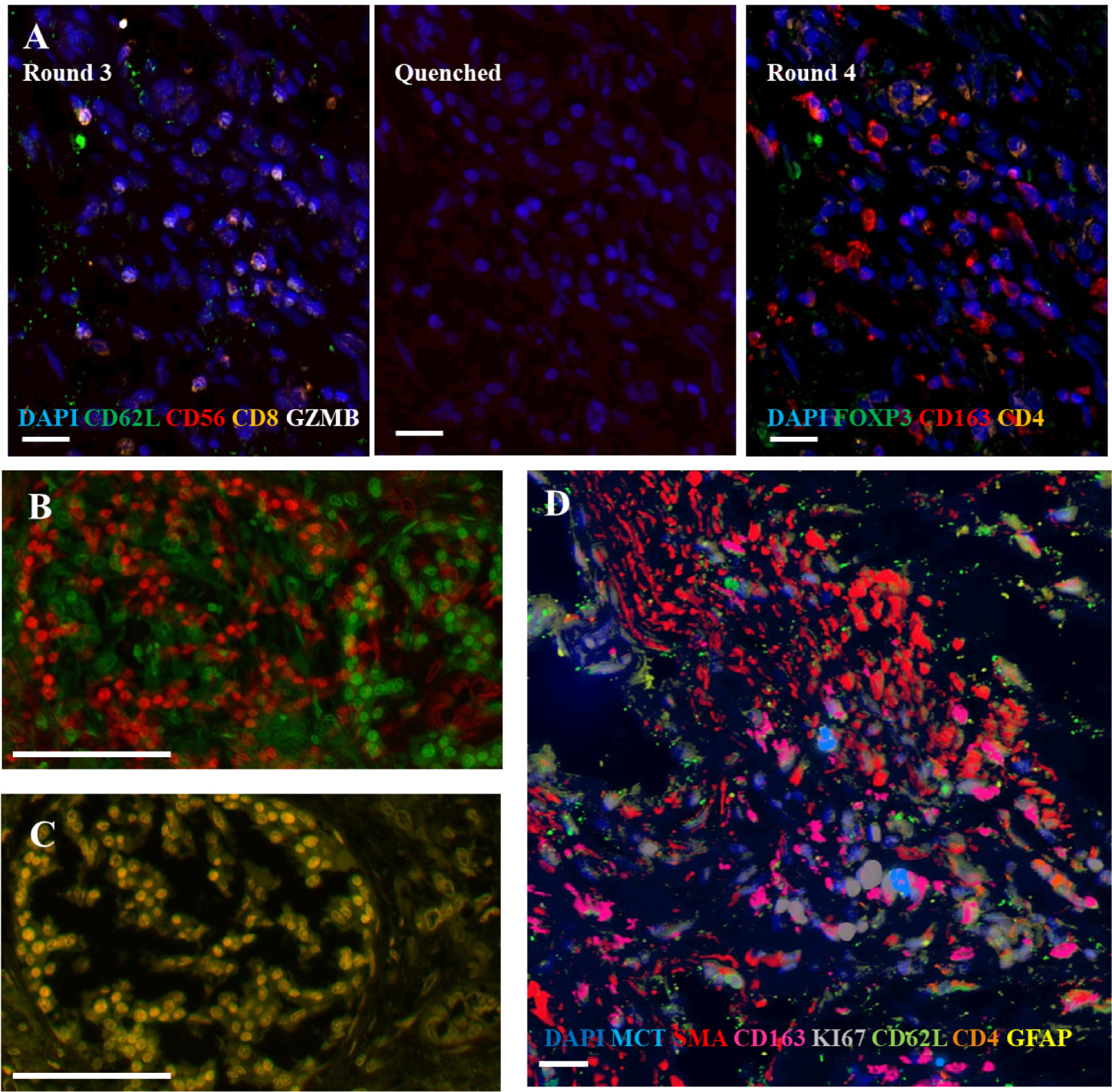
Representative images from cyclic multiplexed-immunofluorescence (MxIF) labeling of a primary PDAC tumor with image registration. A) Images showing distinct marker distributions in the same area across staining round 3, quenching and re-scanning at the same intensity, and re-staining round 4. Scale bar = 20 µm. B) Unaligned image of DAPI staining of the same area from round 1 (green) and 9 (red) . C) Alignment (overlaid yellow) of round 1 and 9 DAPI using image registration software. Scale bar = 100 µm. D) Aligned composite image showing a subset of markers from different rounds of the MxIF panel. Scale bar = 20 µm.
