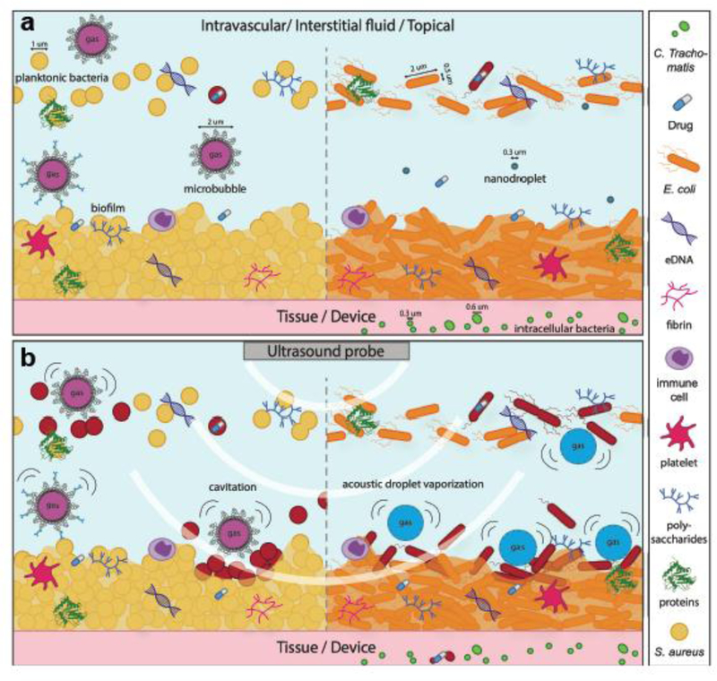
Figure 1.
Concept of sonobactericide (not drawn to scale). a) potential infection environments before ultrasound. Sizing of bacteria and cavitation nuclei are denoted with a line and an arrow on each end. b) ultrasound application where upon cavitating microbubbles and activated nanodroplets disrupt bacteria and biofilm composition. Bacteria that have become red in b) are considered dead or to have compromised membranes due to the effects from ultrasound and cavitating nuclei.

