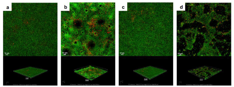
Figure 4.
Confocal laser scanning micrographs of in vitro, propidium iodide (red) stained, Pseudomonas aeruginosa PAO1:gfp-2 biofilms following treatment with a) nothing (control), b) ultrasound and Definity® (microbubble), c) gentamicin (antibiotic) alone, and d) gentamicin, ultrasound, and Definity®. The top panel is the top-down maximum intensity projection and the bottom panel is the corresponding three-dimensional volume rendering. Ultrasound parameters were 0.5 MHz at 1.1 MPa peak negative pressure with a 16-cycle tone burst and pulse repetition frequency of 1 kHz for 5 minutes (reprinted with permission from Ronan et al. (2016)).

