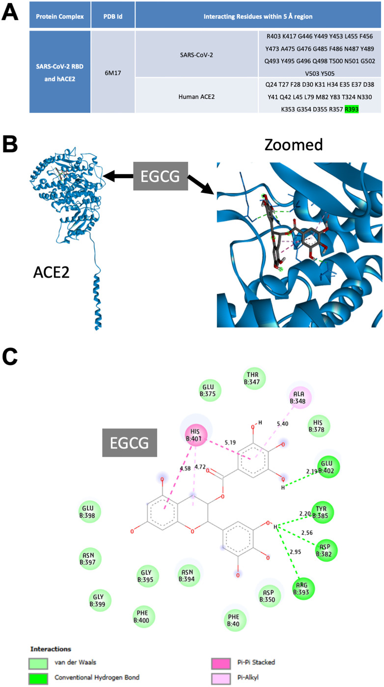
Fig 3. Docking interaction of epigallocatechin gallate (EGCG) with human angiotensin-converting enzyme 2 (ACE2).
(A) Interacting residues determined within a 5-Å region at the interface of the severe acute respiratory syndrome coronavirus 2 (SARS-CoV-2) receptor-binding domain (RBD) and human ACE2 from their crystal structure (6M17). (B & C) Molecular docking of human ACE2 and EGCG. (B) Three-dimensional representation of EGCG binding with the human ACE2 peptidase domain (left) and magnified image (right). (C) Two-dimensional interaction analysis of ACE2 and EGCG, highlighting the ACE2 residues that interact with EGCG and the types and lengths of their bonds. Residues are color coded according to the type of interaction: dark green, conventional hydrogen bonds; light green, van der Waals forces; dark pink, Pi–Pi stacked interactions; and light pink, Pi–alkyl interactions.

