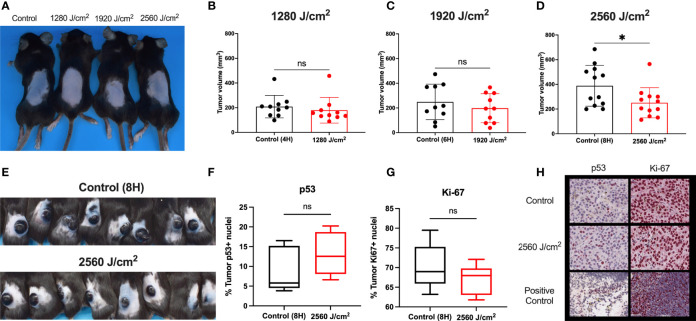
Figure 5.
RL safety and efficacy. (A) C57BL/6 mice without tumors were treated with RL at 1280, 1920, and 2560 J/cm2 for 15 days (n=3). Mice had no increase in rectal temperature, and the skin was non-inflamed and non-erythematous compared to non-RL treated mice. (B) C57BL/6 Mice were injected with 3 x 10⁵ melanoma cells and irradiated daily with 1280 J/cm2 (n=10), (C) 1920 J/cm2 (n=10), and (D) 2650 J/cm2 RL (n=12). Volume was calculated using the formula Volume = 0.52 x length x width x depth. (E) Representative mice with tumors (n=8) in the control group and 2560 J/cm2 RL group on day 15. (F) Quantification of p53+ (n=5) and (G) Ki-67+ (n=5) staining nuclei in from control (8H) and 2560 J/cm2 treated tumors. Quantification of staining was performed using Indica HALO software. (H) Representative images of p53+ and Ki-67+ staining in tumors. Cephalic (embryonic day 14) and spleen positive control sections are provided for p53 and Ki-67. Excised tumor volumes and IHC staining intensity for RL-treated mice were compared to matched controls by a two-tailed T-test (p<0.05). *denotes p<0.05. ns denotes not significant.

